Our Approach to 3D Modeling
Make an Appointment
Our team is here to help you schedule an appointment with the specialists you need. For referring providers, we accept referrals from across the region and nationally for complex cases.
Our team creates patient-specific 3D models using advanced segmentation, rendering, and 3D printing tools. These models support a wide range of congenital heart conditions, including:
- Double-outlet right ventricle (DORV)
- Borderline left or right ventricular size
- Complex outflow tract abnormalities
- Aortic arch anomalies (coarctation, interrupted arch, vascular rings)
- Pulmonary artery abnormalities
- Single ventricle pathways (Stage I–III palliation)
- Anomalous coronary arteries (AAOCA)
- Airway compression associated with vascular anomalies
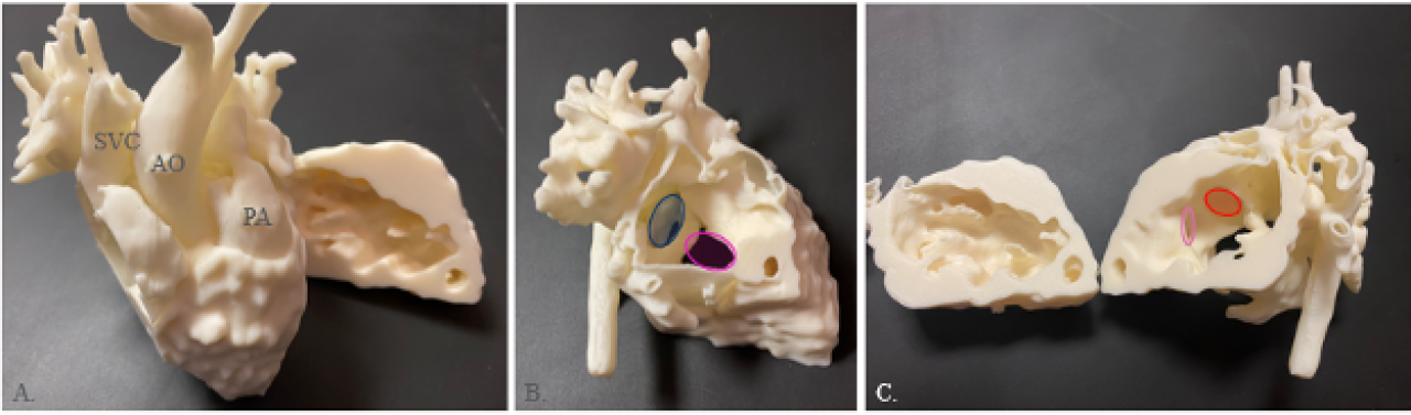
3D Printed model of a patient with double inlet left ventricle.
How 3D Modeling Works: Our Process
Our workflow combines state-of-the-art imaging, engineering, and expert review.

- Acquisition - High-resolution echocardiography, cardiac CT, or cardiac MRI provides the foundational data.
- Segmentation & Model Creation - Specialized software is used by trained engineers and imagers to build an accurate, patient-specific 3D model.
- Multidisciplinary Review & Planning - Cardiologists, surgeons and engineers review the model together to determine the safest and most effective treatment plan.
- Clinical Application - 3D models are used in the operating room, cath lab, clinic discussions with families, and educational settings.