Cardiovascular Imaging
Make an Appointment (Echocardography)
Make an Appointment (Nuclear Cardiology)
Columbia University Medical Center is home to one of the largest and most advanced adult cardiovascular imaging programs in the world and the largest such program in the northeast. Each year, we perform more than 40,000 detailed imaging studies of the heart and vascular system. We are nationally recognized for noninvasive cardiovascular imaging, having pioneered many of the technologies we use today. In 2003, we opened our vascular medicine program, one of the first programs of its kind in the New York region.
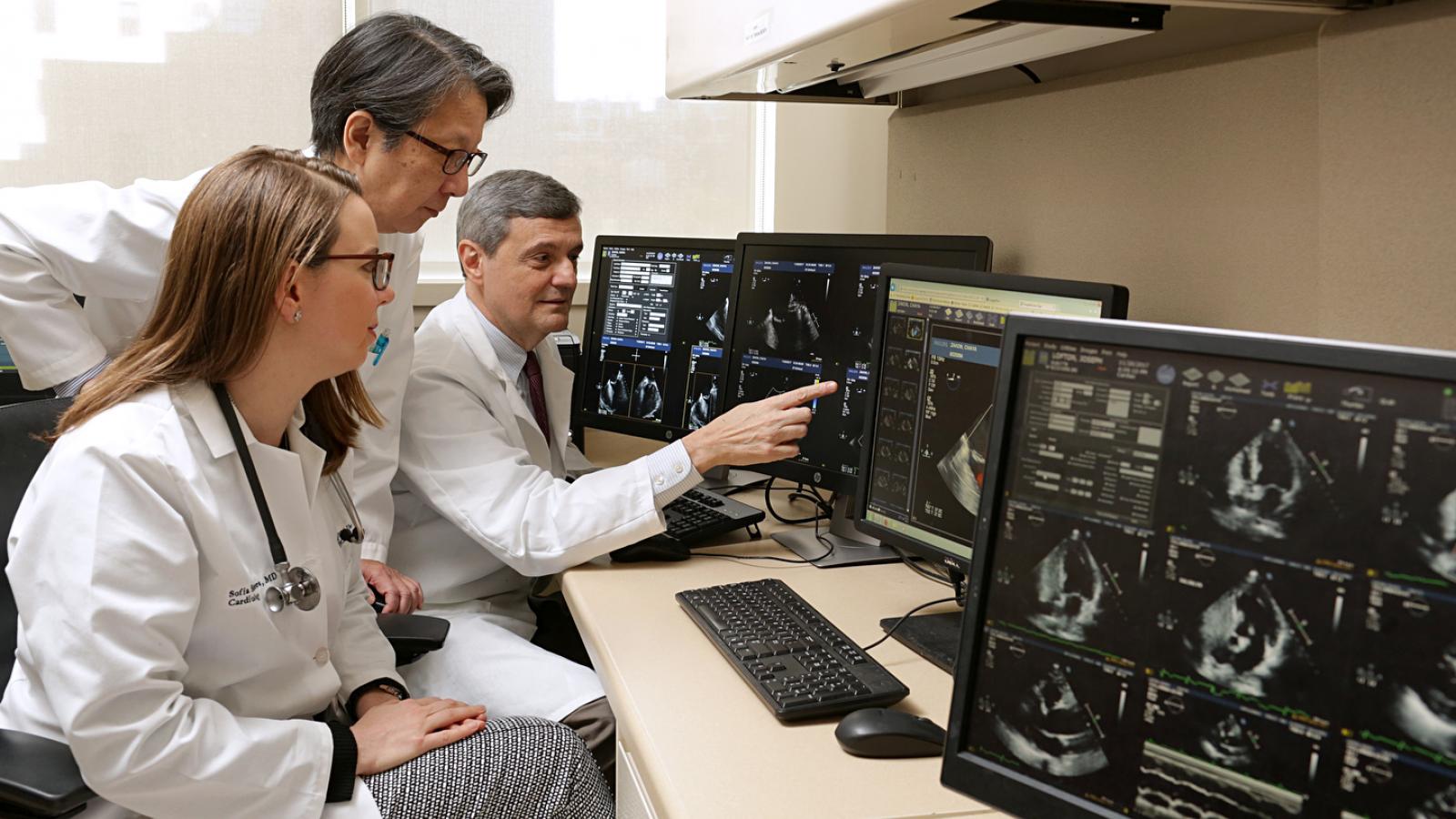
Our physicians are cardiologists and other specialists with advanced skills and specialty training in cardiovascular imaging. Together, our doctors comprise a premier group of imaging experts, each with a particular focus in the fields of echocardiography, nuclear cardiology, and vascular ultrasound. Areas of expertise include valve disease, cardioembolic stroke, percutaneous valve procedures, heart transplantation, ventricular assist devices, Cardiac SPECT, and cardiac PET imaging.
We use echocardiography, vascular ultrasound, and cardiovascular nuclear imaging for the noninvasive diagnosis of many disorders of the heart and blood vessels, including:
- Atherosclerosis
- Cardiac tumors and masses
- Cardiomyopathies
- Carotid artery disease
- Congenital heart disease
- Coronary artery disease
- Deep venous thrombosis
- Diseases of the aorta
- Heart failure
- Renal artery disease
- Valvular heart disease
- Vasculitis
State-of-the-art Imaging Services
Echocardiography
- 2D and 3D transthoracic echocardiography
- 2D and 3D transesophageal echocardiography
- Exercise and pharmacologic stress testing
- LV strain imaging
Nuclear
- Exercise and pharmacologic stress SPECT and PET myocardial perfusion imaging
- Myocardial viability imaging by SPECT and PET
- Cardiac Amyloid imaging by SPECT and PET
- Cardiac inflammation (focal or diffuse), endocarditis, device infection imaging by cardiac PET
- Quantitative coronary flow assessment by cardiac PET
- Coronary Artery Calcium Score
Vascular
- Carotid duplex ultrasound
- Non-invasive flow studies
- Upper and lower extremity arterial and venous ultrasound
- Abdominal aorta and renal vascular ultrasound
Cardiac CT
- Detection of coronary artery disease
- Assessment of suspected anomalous coronary artery
- CT angiography for annular and vascular evaluation prior to transcatheter aortic valve replacement
Cardiovascular MRI
- Assessment of ventricular function, size, wall thickness, mass and valvular pathology
- Assessment of congenital heart disease, cardiac masses, and pericardial disease
- As part of diagnostic evaluation for arrhythmogenic right ventricular cardiomyopathy
- Contrast study for evaluation of myocardial viability, infarction,
- cardiomyopathies, myocarditis, and diseases of the great vessels